
|
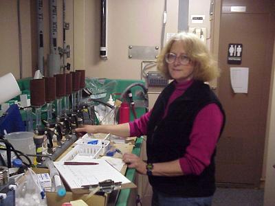
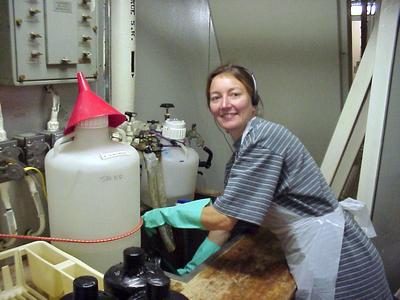
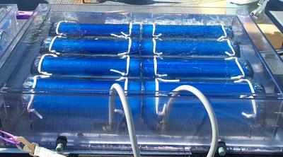
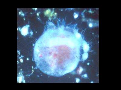
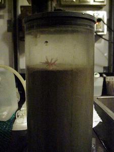
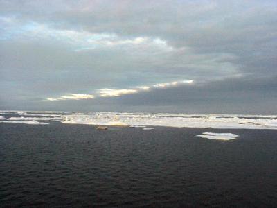
8 August, 2002Yesterday (I'm writing this on August 9) seemed like a very long day! Things started well when I got up early to write the journal for the 7th. The journal was almost done when my computer, which had been acting strangely for a few days, suddenly froze up. After shutting it down and restarting it, it closed itself down and gave me a screen-wide error message. After I shut it down and restarted it one more time, it shut itself down again and would not allow me to re-boot (start it up again)! Knowing that all my journals and photos were saved on the hard drive, I was very nervous! Two wonderful people, Steve Roberts and Sean Kuhn (I'll tell you about each of them in an upcoming journal), spent several hours working on it and they managed to get all my files backed up onto the ship's computer. My own computer is running again, but doing strange things periodically, so I will continue to back up all my files daily using the ship's computer. The lesson for today is - always back up your important files on a regular basis. Our sediment sampling started at 4:45 PM and we wrapped up our work at 1:45 AM. It was a difficult station because the station is right on the slope which drops off sharply to the basin. Whenever the ship is stopped for sampling it drifts. Usually that isn't a problem because the depths don't change that quickly. But, due to our position on the slope we had to re-position several times to be certain we were sampling at the correct depth of 500 meters. In addition, the mud was soft, making it difficult to get a good core sample. Jackie is not one to give up, and we kept trying until she had the samples she needed. At one point in the evening we stopped for yet another amazing organism in one of Jackie's samples. You often see pictures of coral reefs with brightly colored anemones attached to the coral, waving their tentacles into the water above them. Jackie had found a beautiful, orange colored anemone in the sediments at 500 meters! One other note - around midnight, the skies began to clear for the first time in several days. As the sun broke through the clouds the ice seemed to glow with light; it was spectacular! After a few hours rest, I got up this morning to talk with Ev Sherr and Jennifer Crain. Ev is a professor of oceanography at Oregon State University in Corvallis, Oregon where Jennifer is a science technician. We all admire Jennifer because she is one of a handful of people who were on the SBI spring cruise and who, after a brief three weeks at home, returned for the SBI summer cruise. Remember that each cruise is 40 days long! Ev and Jennifer are working closely with Carin Ashjian, Bob Campbell (he came on board about a week ago) and Stephane Plourde to study the structure of the plankton food web. If you check the journal from July 27, you'll find the diagram of who eats what among the plankton. Remember that phytoplankton are the plant plankton and zooplankton are the animal plankton. Carin and Bob are studying the grazing rates of the mesozooplankton (larger ones), and Ev and Jennifer are studying the microzooplankton (micro = tiny). Both types of zooplankton eat the phytoplankton, and the mesozooplankton also eat the microzooplankton (check the diagram in the July 27 journal). The microzooplankton are extremely small and most are in a kingdom of unicellular organisms called Protista. If you've ever looked at pond water under a microscope, you've probably seen examples of protists swimming around. While Carin and Bob are looking at the grazing rates of the mesozooplankton, Ev and Jenn are looking at the grazing impact of protists (microzooplankton) on phytoplankton. In order to do this, they take large amounts of water from one depth. They choose a depth that is in the euphotic zone (light reaches there) where they are certain that there are lots of phytoplankton. They concentrate on the innermost stations (shelf) and outermost stations (basin) in each transect. Once they have their water, Ev and Jennifer dilute it several times with filtered water before they incubate it (put it in an environment where things can grow naturally) in two large tanks on the bow of the ship. They do this to make it more difficult for the grazers (protists in this case) to feed on the phytoplankton. Each incubation tank contains eight cylinders and each cylinder contains 2 large bottles of water. Each cylinder is wrapped in a special plastic to mirror in situ (natural) light levels. The incubation takes three days because the water is so cold that life processes are very slow. Both before and after incubation, Ev and Jennifer sample the water for chlorophyll. Since phytoplankton contain chlorophyll, these chlorophyll measurements give them an idea of the change in the numbers of phytoplankton over the three days. Using that information, they can calculate the growth rate of phytoplankton both with and without grazing (other organisms eating them). For the last part of their work, Ev and Jennifer filter their water to remove the protists and use a special stain in order to view them using epifluorescence microscopy. That means they use a specialized microscope where the light comes from the top (epi). Stop and think about the big picture for just a minute. Phytoplankton are the base of the ocean food web. Both micro and mesozooplankton eat them, and mesozooplankton also eat the microzooplankton. Now think about an even bigger picture. Zooplankton are eaten by fish which are eaten by seals which are eaten by polar bears! Maybe now you can appreciate why it is so important to understand the ocean food web (who eats what) at ALL levels.
Contact the TEA in the field at . If you cannot connect through your browser, copy the TEA's e-mail address in the "To:" line of your favorite e-mail package. |