
 | |||||||||||||||||||||
|
|
Journals 2009/2010Jacqui Smoler
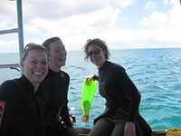
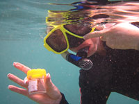
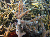
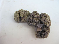
November 12, 2009 HI09-006, depth 1-2 m, 70 min Today I went out on the Chromis, an inflatable boat, with Cruise Leader Aaron and a team of marine biologists including Abby, Phil, Rob, Sea, Kareen and Francois. The boat is named after the Bluegreen Damsel fish, Chromis viridis, which is a common inhabitant of the local reef habitat. We headed out on a short fifteen minute trip to First Point, North Heron Reef to collect specimens from a back-reef habitat. After a few initial problems with my perished snorkel, Aaron declared that it was unfit for future use. I was however fortunate to be able to borrow Kareen's snorkel as she was diving on this occasion. With the new snorkel fitted snugly to my mask I headed out to the reef some 50 metres from the boat with Abby who was keen to collect some algae. I decided that I would help her find some algal specimens as phycology was my area of expertise during my university days. While surveying the reef I also collected an unusual specimen of coral which looked like broccoli. Abby had an underwater camera and took some photos of the specimens and a few of me in snorkeling mode. As I snorkeled along the reef I was mesmerized by the dazzling colour, beauty and variety of corals, fish and bryozoans. Nature really is beautiful!
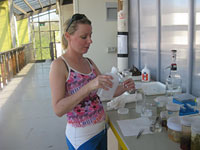
In the afternoon most of the biologists analysed the specimens that they had collected earlier in the day during either a dive or snorkeling expedition. I offered to assist Dr Monika Schlacher prepare the soft coral specimens for both DNA and microscopic analysis. Using forceps and a scalpel I dissected tiny piece of tissues about 3-4 mm in length from each specimen and placed one piece in an Eppendorf tube. Each tube was then topped up with 100% ethanol and the lid was sealed in preparation for DNA analysis by one of the genetic scientists. The next task involved placing even smaller samples of tissue in another set of Eppendorf tubes for sclerite analysis. Sclerites are polycrystalline aggregates of calcite (calcium carbonate), which come in all sorts of shapes and are used by taxonomists to assist them in the classification of soft coral species. After placing the soft coral tissue into the tubes, the tubes were then half-filled with bleach to soften the tissue and release the harder sclerites, which then sank to the bottom of the tubes. Each of the 14 tubes then had to be rinsed to prevent the bleach from forming crystals on the microscope slides. This involved removing the bleach by tipping the contents into a waste beaker and then pipetting distilled water into the tubes, a process which had to be repeated three times to ensure that the bleach had been thoroughly removed. The final process involved placing a pipette into each tube and slowly drawing up a small sample of the sclerites and placing three drops at the end of a labeled microscope slide for taxonomic analysis. I was particularly keen to analyse the broccoli-like specimen that I had collected earlier in the day. Together, Monika and I observed its sclerites and decided that it was in the genus Nephthya sp. We analysed at least six other specimens and Monika recorded the species in a notebook for future reference. Monika was particularly interested in an unusual specimen that she had never seen before and after documenting its sclerite shape and photographing it she decided to send her findings to an expert colleague for further analysis. She later found out that it belonged to a related group of corals called the sea pens.
|
||||||||||||||||||||
